
Two weeks after birth he developed an IH on the right chest and was referred to our hospital for oral treatment. The complication of treatments (hypotension, bradycardia, hypoglycemia, hyperkalemia, wheezing) does not occur. We calculated the IH surface area and volume at the start of treatment, and 1, 3, and 6 months after starting medication and followed up until three years old (1 case was lost to follow-up 1 month after starting medication and 2 cases were lost to follow-up 3 months after starting medication). The study cohort consisted of 2 boys and 8 girls ( Table 1). The average age at starting medication was 3.5 months. Careful consideration was paid to protecting the privacy and personal information of the subjects. The purpose of the study was explained in detail and the participants’ parents voluntarily submitted a signed, written informed consent.
#VECTRA 3D IMAGING AT HOME CODE#
This study was reviewed and approved by the Ethical Review Board of Keio University before patient enrollment and data collection (approval code No. The IH lesion outlines were manually traced to automatically calculate the surface area and volume.
#VECTRA 3D IMAGING AT HOME SOFTWARE#
3D reconstruction was generated using the attachment software for the 3D camera. Because the 3D camera system is designed to take pictures of people, the shooting time is 0.002 seconds, the color image is highly accurate, and background noise is suppressed.

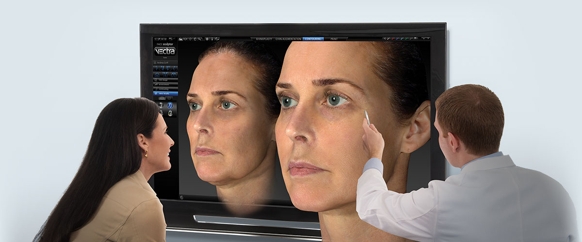
The Vectra H1 is a handy 3D camera system the images are easy to analyze and the volume measurements are easy to obtain. 3D photographs of the IHs were taken using a 3D camera (Vectra ® H1) before starting the medication, and 1, 3, and 6 months after starting the medication).

We slowly tapered the 2 mg/kg per day dose after 6 months and stop the medication. After discharge from the hospital, we followed the patients in the outpatient clinic. We initiated propranolol treatment in the hospital at a dose of 1 mg/kg per day and increased the dose to 2 mg/kg per day. Baseline screening by pediatric cardiologists included blood pressure, heart rate, hematologic examination, an echocardiogram, and an electrocardiogram. Oral propranolol treatment was provided for those who were expected to cause serious dysfunction such as respiratory and visual function or expected to have surgical resection after spontaneous regression, and for IHs in areas likely to be wedge-shaped (nose, lips, auricle, intercostal, etc.). All IHs were in the proliferative phase and 3 months after. This study included 10 patients treated with oral propranolol treatment from 2015-2016 in our hospital before propranolol treatment was covered by insurance in Japan. In this study, we determined whether or not 3D photography is a useful tool for measuring the surface area and volume of IHs over time during treatment. To overcome the shortcomings, we used a 3D camera (Vectra ® H1 Canfield Scientific, Inc., NJ, USA) to measure quantitative changes in IHs. Because the indication was limited by expense and radiation exposure, there were many patients with superficial lesions who were ineligible, but there were several patients who were eligible, such as those with an IH in the orbital space or huge and deep IHs. Although digital cameras are very simple, changes in color tones occur over time, lesions are evaluated in two dimensions, and changes in the thickness of IHs are difficult to evaluate. To evaluate the effectiveness of propranolol oral administration, we first measured quantitative changes in IHs by digital camera and CT or MRI. Before national insurance approval, we offered this treatment in our hospital after review and approved by the Ethical Review Board of Keio University School of Medicine (approval code No.

The oral administration of propranolol for treatment of IH has been covered by national insurance in Japan since 2017. Propranolol is now the recommended first-line oral therapy for IH. The effectiveness of oral propranolol in the treatment of IHs was first reported in Europe in 2008, and a global randomized control study was conducted. Because it takes a long time for the color of IHs to fade and residual lesions have a wrinkle-like appearance, it is sometimes necessary to surgically remove scars. After the proliferative phase, the color of most IHs fades and the size decreases. Infantile hemangiomas (IHs) rapidly increase in size from birth until 5 months of age.


 0 kommentar(er)
